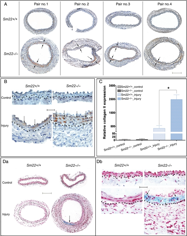
Figure 1.
Enhanced type II collagen in Sm22−/− mice 2 weeks after carotid denudation. Expression of type II collagen was evaluated by IHC. (A) Injured carotids from four Sm22−/− mice and their wild-type littermates at 100× magnification are shown. (B) Both injured carotids and non-injured controls at 400× magnification. Representative brown signals were indicated by the arrows. (C) Quantification of positive signals from images at 100× magnification in the media of carotids from five Sm22−/− and their littermates Sm22+/+ mice. (D) Alcian blue staining of carotids at 100× (Da) and at 400× magnification (Db). Blue signals, Alcian blue; red signals, nuclear fast red. Bar in (A and Da), 100 µm; bar in (B and Db), 20 µm. Dashed lines demarcated the border between media and adventitia. Values are means ± SE. The asterisk indicates P < 0.05 vs. Sm22+/+ mice.

