Figure 1.
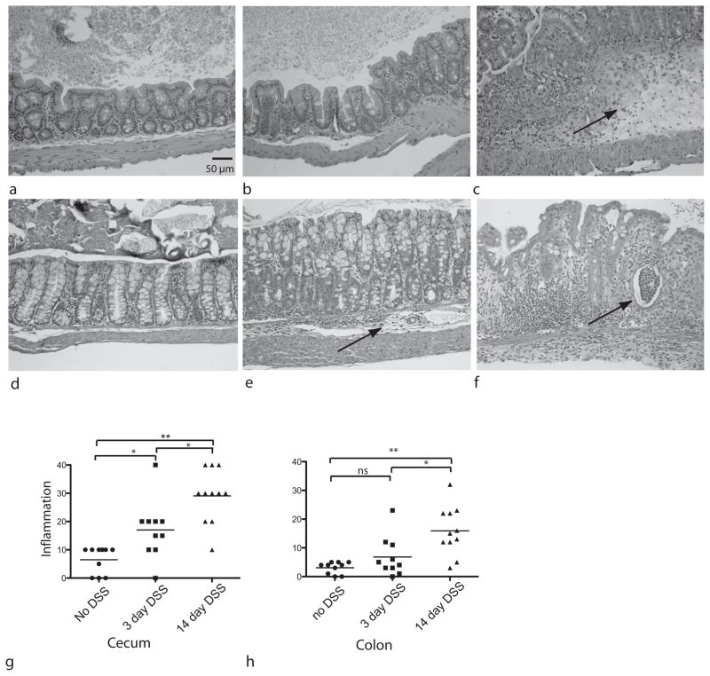
Histopathology in DSS-treated mice. Hemotoxylin and eosin (H&E) stained sections were prepared from the cecum of (a) untreated control mice (b) mice after 3 days of DSS treatment and (c) after 14 days of DSS. Arrow indicates submucosl edema. H&E sections were also prepared from colon samples of (d) untreated controls (e) mice after 3 days of DSS treatment (arrow points to inflammation) and (f) animals after 14 days of DSS (arrow shows abscess). Histopathologic scores were calculated for sections from all 31 animals for (g) cecum and (h) colon sections. Statistical analysis done was Kruskal-Wallis test. *p< 0.05 **p<0.001. Initial magnification 40X

