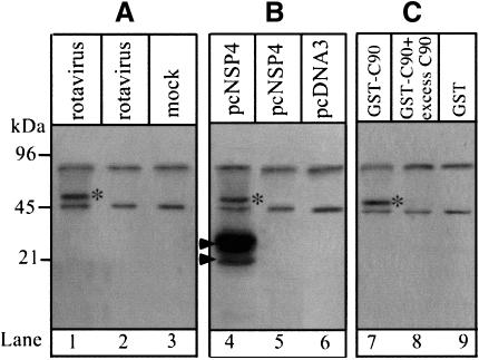
Fig. 1. NSP4 interacts with a 50 kDa cellular protein. (A) Immuno precipitation of labelled proteins from MA104 cells. Cells were 35S-labelled for 6 h and chased for 1 h prior to infection (lanes 1 and 2) or mock infection (lane 3) with SA11 rotavirus. Cells were harvested at 7 h post-infection and lysates immunoprecipitated with anti-NSP4 mAb (lanes 1 and 3) or pre-immune serum (lane 2). (B) Immunoprecipitation of labelled cellular proteins from transfected Cos-7 cells. Cells were transfected with pcNSP4 (lanes 4 and 5) or pCDNA3.1 (lane 6) and 35S-labelled for 6 h commencing 40 h after transfection. Lysates were immunoprecipitated with anti-NSP4 mAb (lanes 4 and 6) or pre-immune serum (lane 5). Arrows indicate the positions of glycosylated (upper) and unglycosylated (lower) NSP4. (C) Pull-down assay using the C-terminal 90 amino acids of NSP4 (C90) fused to GST as bait. The procedure is described in Materials and methods. The specificity of the interaction was tested by addition of excess C90 (100 µg/ml) (lane 8). Proteins were resolved by 12% SDS–PAGE and visualized by autoradiography. *, the 50 kDa cellular protein identified in each experiment.

An official website of the United States government
Here's how you know
Official websites use .gov
A
.gov website belongs to an official
government organization in the United States.
Secure .gov websites use HTTPS
A lock (
) or https:// means you've safely
connected to the .gov website. Share sensitive
information only on official, secure websites.
