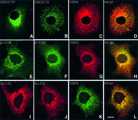
Fig. 8. Distribution of ERGIC53, β-COP and Sec13p in cells expressing NSP4. Cos-7 cells were transiently transfected with pcNSP4 (B–D, F–H and J–L) or mock transfected (A, E and I) and grown for 48 h. Cells were fixed, permeabilized and double-labelled with a polyclonal antibody against NSP4 and a monoclonal antibody against either ERGIC53 (A–D), β-COP (E–H) or Sec13p (I–L) prior to analysis by confocal laser scanning microscopy. The scale bar represents 10 µm.

An official website of the United States government
Here's how you know
Official websites use .gov
A
.gov website belongs to an official
government organization in the United States.
Secure .gov websites use HTTPS
A lock (
) or https:// means you've safely
connected to the .gov website. Share sensitive
information only on official, secure websites.
