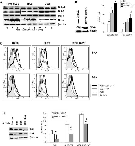
Figure 4. Effect of GSI/ABT-737 combination on apoptosis of MM cell is mediated through activation of Bak and Bax.
(A) MM H929, RPMI 8226, or U266 cells were treated with GSI for 48 hrs. After that time cells were collected and the level of indicated proteins was determined by Western blotting. Vertical lines separate part of the membranes that were developed using maximum strength enhanced chemiluminescence. (B, D) MM 8226 cells were transfected with siRNA specific for Noxa, bak, bax, or control non-targeting siRNA. After overnight incubation cells were treated with GSI, ABT-737 or drug combination. Western blotting with antibodies specific for Noxa (B) or Bak and Bax (D) was performed. Apoptosis in different treatment groups of cells was detected by Annexin V/DAPI staining using LSR II flow cytometer (BD). Shown are combined results of three independent experiments performed in duplicates. (B) * - statistically significant difference (p<0.05) between indicated group and GSI+ABT-737 group; # - statistically significant difference (p<0.05) between cells transfected with control or Noxa siRNA and treated with combination of GSI and ABT-737. (D) * - statistically significant difference (p<0.05) between cells transfected with control or Bak and Bax siRNA. (C) MM U266, H929, or RPMI 8226 cells were cultured for 48 hrs with or without GSI, ABT-737, or combination thereof. Cells were then collected and the level of Bax and Bak activation was evaluated by flow cytometry as described in the Methods section. Mean fluorescence intensities were determined and compared between cells treated with ABT-737, GSI, of combination thereof.

