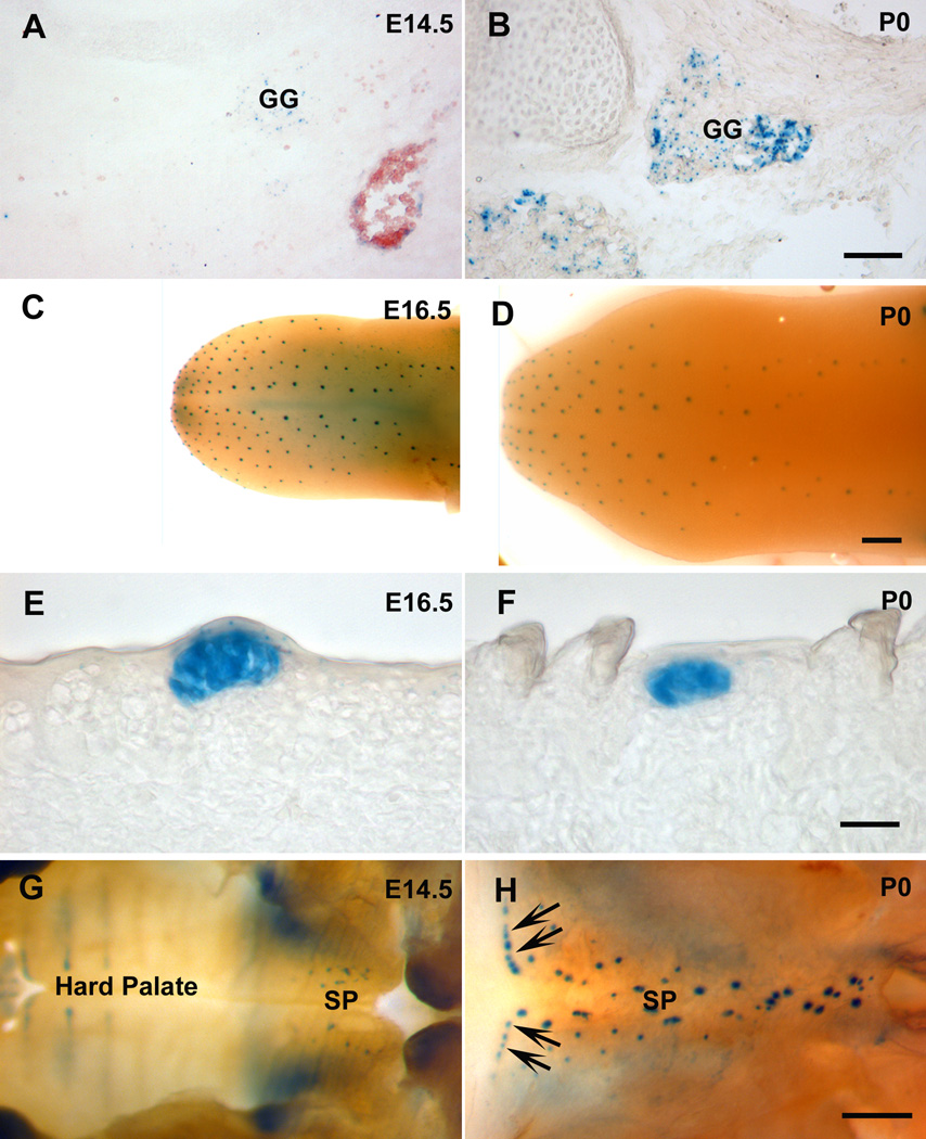
Figure 6. The changes of β-gal staining in BdnfLacZ/+ mice are consistent with the changes revealed by RT-PCR.
β-gal was detected in geniculate ganglion (GG) at both E14.5 (A) and birth (B), but much stronger staining was observed at birth. Whole mount β-gal staining of the tongue demonstrated that the distribution pattern of β-gal-positive spots was similar between E16.5 (C) and birth (D), but the intensity of staining for β-gal in E16.5 tongue was stronger than that at birth (E and F). β-gal-positive signals were found in soft palate (SP) at both E14.5 (G) and birth (H), but more at birth, especially in the region corresponding to the Geschmacksstreifen (arrows). Scale bar is 100 µm for A and B; 1000 µm for C and D; 20 µm for E and F; 400 µm for G and H.

