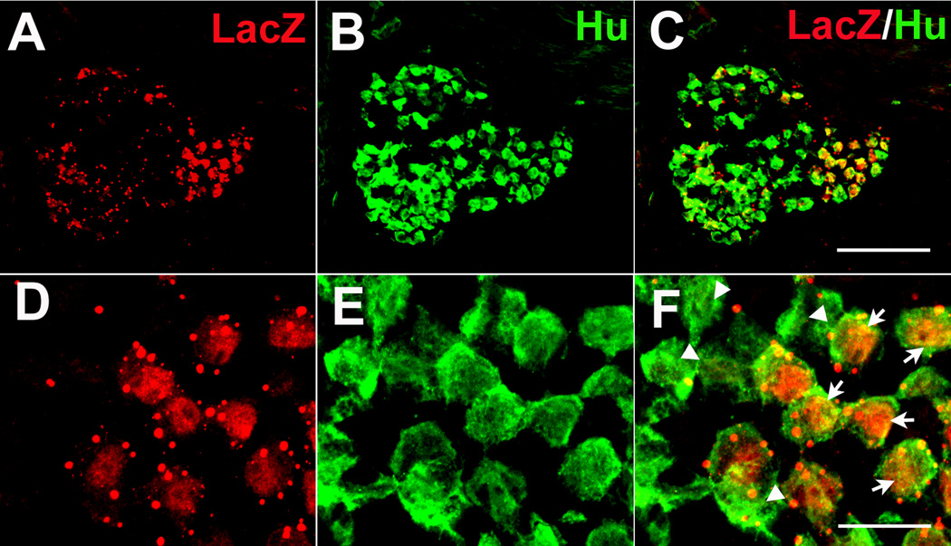
Figure 7. BDNF is expressed in the geniculate ganglion neurons at birth.
Double label immunohistochemistry at birth illustrates that BDNF (A: anti-β-gal) is expressed in some neurons (B: anti-Hu) but not all (C). This can be seen more clearly at higher magnification (D, E, F). The arrows indicate cells that co-express β-gal and Hu. Arrowheads indicate β-gal negative neurons. Scale bar in C, 100 µm, applies for A, B and C; Scale bar in F, 20 µm applies for D, E and F.

