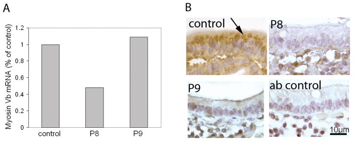
Figure 2.
Real-time PCR detection of myosin Vb mRNA and immunohistochemical labeling of myosin Vb. A) RNA was extracted from small intestinal biopsies of MVID patient 8 and 9 (designated P8 and P9) as described in Materials and methods, and relative myosin Vb mRNA levels was determined with real-time PCR. B) small intestinal biopsies of MVID patient 8 and 9 and age-matched controls were labeled with antibodies against human myosin Vb. Negative (non-immune first antibody) control staining are shown. The accumulation of myosin Vb in the apical cytoplasm in control enterocytes (arrows) is lost in MVID enterocytes.

