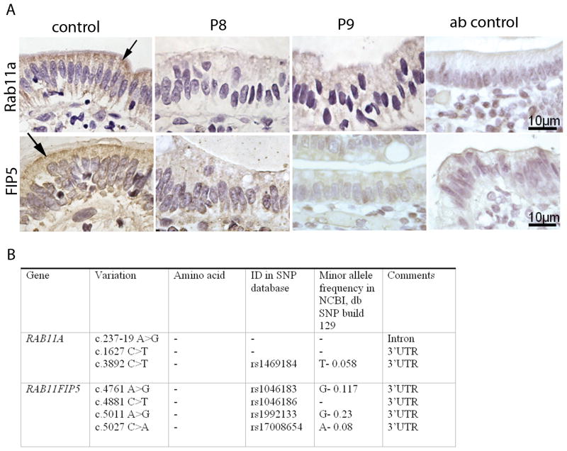
Figure 3.
Immunohistochemical labeling and variant analysis of Rab11a and FIP5(/Rip11). A) The distribution of Rab11a and FIP5(/Rip11) in control and MVID enterocytes of patients 8 and 9 (designated P8 and P9) is shown. Negative (non-immune first antibody) staining is shown (Ab control). Note that the accumulation of Rab11a in the apical cytoplasm of control enterocytes (arrows) is lost in MVID enterocytes. B) Sequence variants in the genes RAB11A and RAB11FIP5.

