
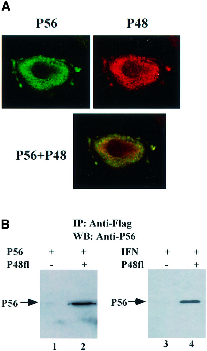
Fig. 2. The interaction of P56 and Int-6/P48 in human cells. (A) Co-localization of P56 and P48 in the cytoplasm. HT1080 cells were transfected with pCMV-P48Fl and, 16 h post-transfection, cells were treated with 1000 U/ml IFN-β for 16 h. A confocal immunofluorescence assay was performed using anti-P56 antibody and anti-Flag antibody. The subcellular locations of P56 (green) and P48 (red) and their co-localization (yellow) are shown. (B) The interaction between both exogenous and endogenous P56 with P48. In lanes 1 and 2, HT1080 cells on a 100 mm plate were transfected with 10 µg of pCMV-P56 alone (lane 1) or co-transfected with 8 µg of pCMV-P56 and 8 µg of pCMV-P48Fl (lane 2). In lanes 3 and 4, cells were transfected with 8 µg of vector alone (lane 3) or pCMV-P48Fl (lane 4) and, 24 h post-transfection, cells were treated with 1000 U/ml IFN-β to induce endogenous P56. After 24 h, cell extracts were made and immunoprecipitation was performed with Flag antibody-conjugated Sepharose beads followed by western blotting with P56 antibody.