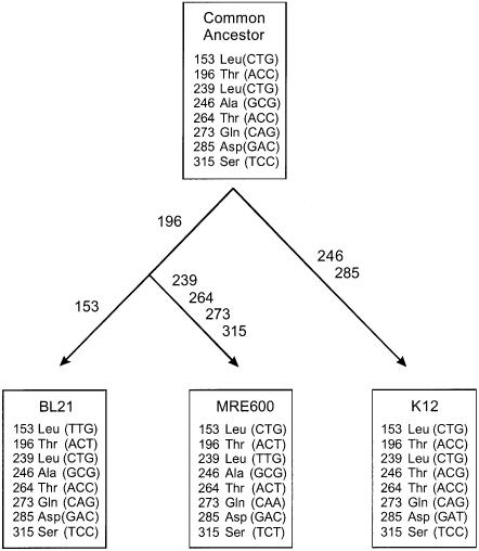
Fig. 1. Codon differences between prfB from different E.coli strains. The lower three boxes show the differences revealed by nucleotide sequencing. The upper box shows the codons likely to be present in a common ancestor of the K12, B and MRE600 strains, based on a comparison of the E.coli sequences and the prfB sequence from S.typhimurium (see text).

An official website of the United States government
Here's how you know
Official websites use .gov
A
.gov website belongs to an official
government organization in the United States.
Secure .gov websites use HTTPS
A lock (
) or https:// means you've safely
connected to the .gov website. Share sensitive
information only on official, secure websites.
