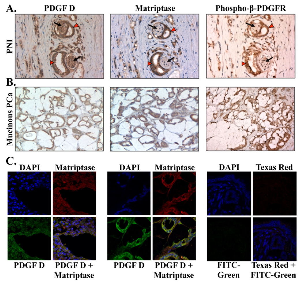
Fig. 6. Expression and co-localization of PDGF-D, matriptase, and phosphorylated-β-PDGFR in PNI and mucinous prostate carcinoma.
A,B. Serial sections of neoplastic glands with perineural invasion (PNI, at 400×) (A) and sections of the mucinous variant of poorly differentiated PCa (mucinous PCa, at 200×) (B) were immunostained with anti-PDGF-D, anti-matriptase, and anti-phospho-β-PDGFR antibodies. Carcinoma and nerve cells are indicated by red arrow heads and black arrows, respectively. C. Immunofluorescence analysis of matriptase (Texas Red) and PDGF-D (FITC-Green) in PCa with Gleason score 8 (left panel) and in a mucinous variant of PCa (middle panel) was performed at 630× magnification. Cell nuclei were stained with DAPI (blue). Yellow in merged panel indicates co-localization of the proteins.

