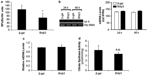
Figure 6.
Bnip3 reduces cellular ATP levels without decreasing the number of mitochondria. (a) At 48 h after infection, cellular ATP levels were measured in Bax/Bak DKO MEFs overexpressing β-gal or Bnip3 (n=6). (b) Mitochondrial DNA content in Bax/Bak DKO MEFs. Shown is a representative gel of PCR for mitochondrial cytochrome b (cyt b) and nuclear 18 s rRNA. Densitometry of cyt b normalized to 18S rRNA (n=3). (c) Real-time quantitative PCR of mtDNA levels at 24 h after infection (n=3). (d) Citrate synthase activity in cells after 48 h of β-gal or Bnip3 overexpression (n=4). Data are mean±S.D. (*P<0.05 compared with β-gal)

