Fig. 4.


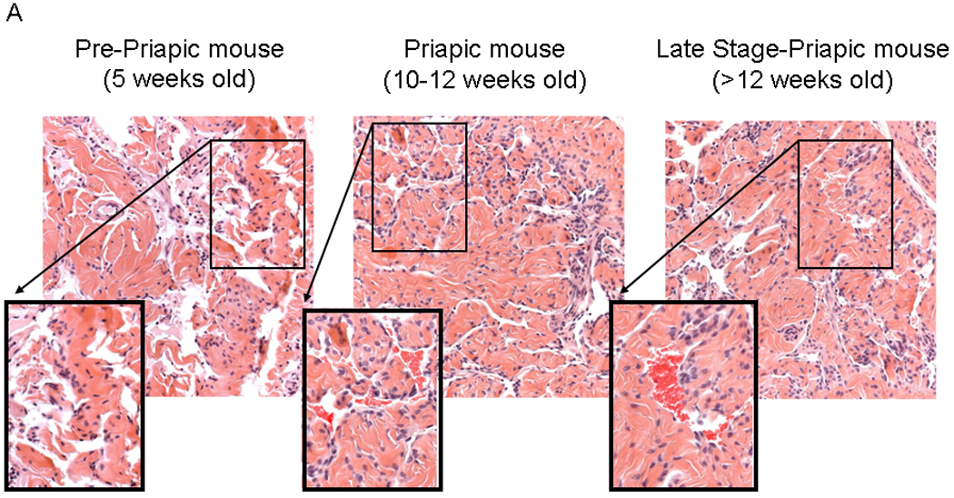
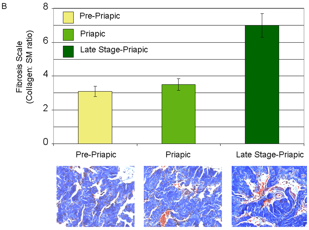
4A. (Used with permission from the American Journal of Physiology, Kanika et al. 9) Histopathology demonstrating the difference between the different life stages of the sickle cell mice. Left panel- pre-priapic mice (Pre-P, there was no evidence of priapism in animals, and histopathology showed no evidence of vascongestion). Middle panel- priapic mice (P, animals were observed to have priapism, and histopathology demonstrates vasocongestion of the corporal tissue). Right panel- late stage-priapic mice (Late stage-P, animals had obvious signs of necrosis of the corporal tissue. Histopathology demonstrated evidence of enlarged spaces with vasocongestion and inflammation of coporal tissue). Sections of each panel are enlarged, and the presence of blood cells within the vascular spaces enhanced to highlight vasocongestion. 4B (Lower Panel) Representative trichrome staining of pene sections from pre-priapic, priapic and late stage priapic sickle cell mice. (Upper Panel) Bar graph demonstrating the ratio of collagen to smooth muscle in the different groups (N=3 animals per group). The late stage-priapic animals had a significant increase in the collagen to smooth muscle ratio compared to the pre-priapic and priapic mice (P<0.05).
