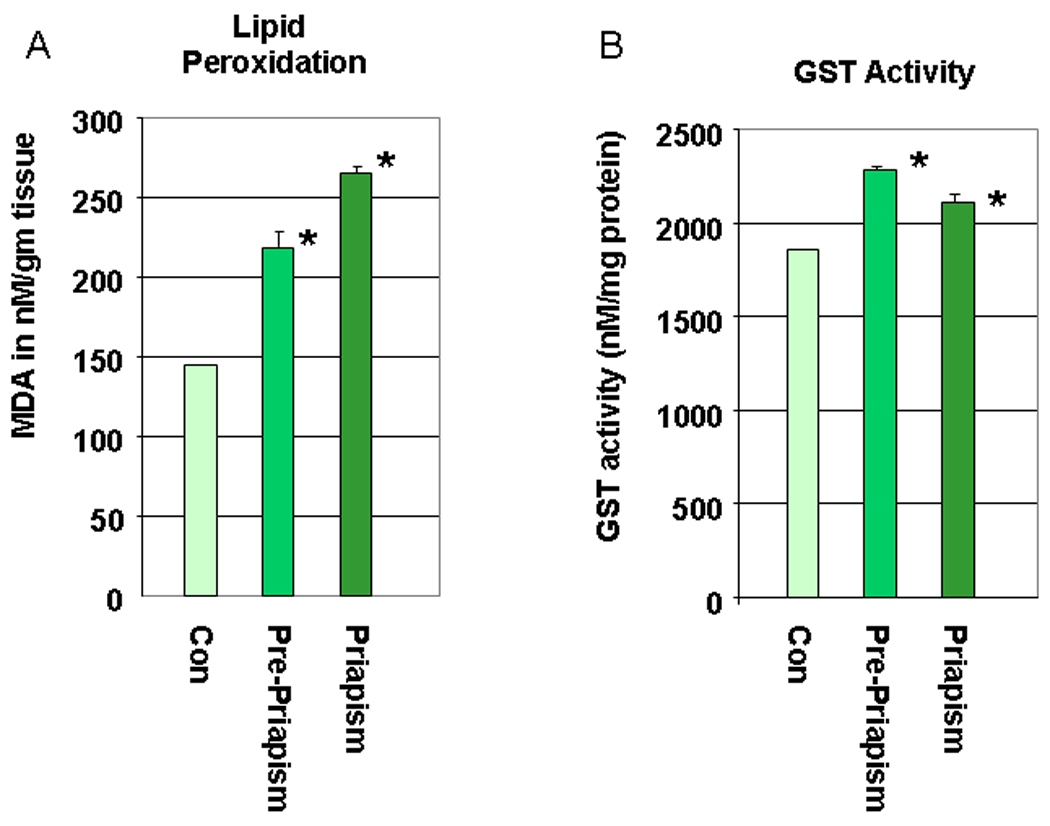
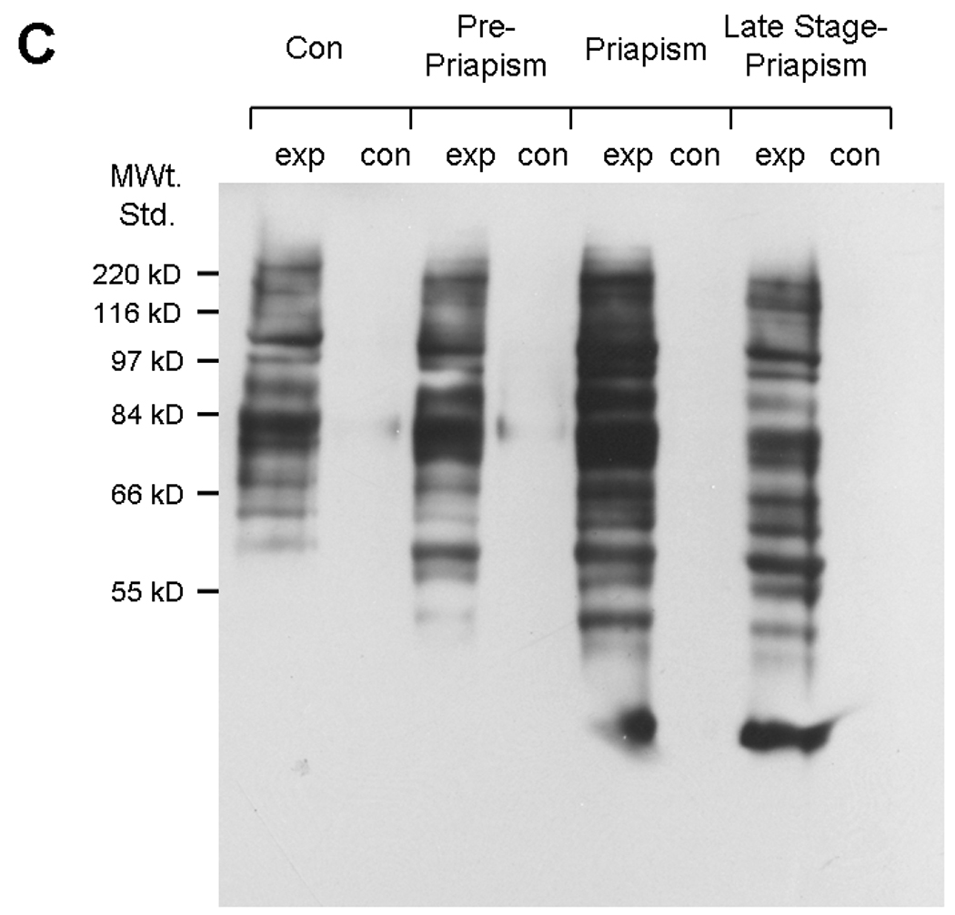
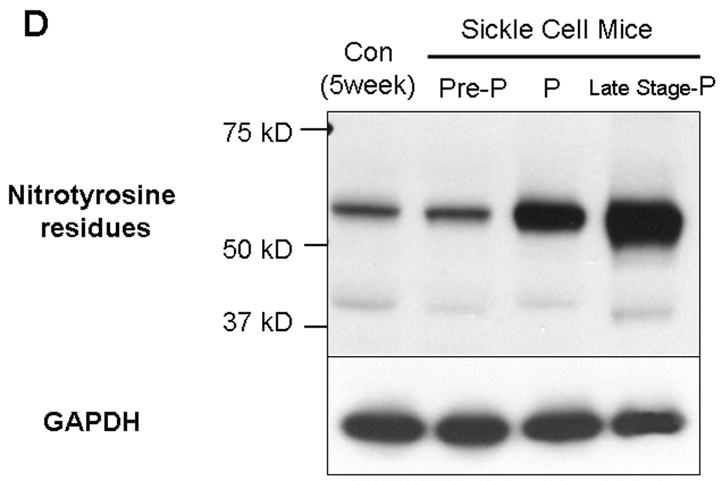
Fig. 5.


5A. Lipid peroxidation was measured by determining malondialdehyde (MDA) in coporal tissues of sickle mice at life stage before the onset of priapism (pre-priapism, with evidence of priapism and in the parent mouse line (C57BL/6). Measurements were performed on three animals in each group in duplicate. * =significant difference from control (P<0.05). 5B. Glutathione S-transferase (GST) activity was also measured in the same animals. * =significant difference from control (P<0.05). 5C. A representative oxyblot performed on protein extracts from the corpora of control mice, or sickle cell mice at different life stages. Protein loading was normalized using a protein assay prior to loading, and confirmed using ponceau staining after transfer to PVDF membranes. The “con” lanes are protein samples that were not derivitized with dinitrophenylhydrazine, and act as negative controls for the assay of samples “exp” that did undergo dinitrophenylhydrazine derivitization. 5D. Representative blot for the global analysis of nitrotyrosine residues in protein extracts from the corpora of control mice, or sickle cell mice at different life stages. Protein loading was normalized using a protein assay prior to loading, and confirmed using ponceau staining after transfer to PVDF membranes.
