
Fig. 2.
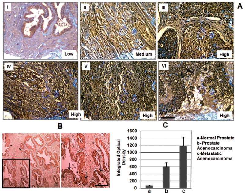
(A) Immunohistochemical analysis of RecQL4 expression in normal (I), malignant (II & III) and metastatic prostate tumor tissues (IV–VI). Prostate tissue panel arrays (PR951 and PR751) purchased from US Biomax Inc., MD, USA were utilized. Malignant tumor samples shown in II (67 year old, Gleason score 7, PSA 37.3 ng/ml) and III (63 year old, Gleason score 10, PSA not determined) illustrate that the RecQL4 expression increases with increasing tumor grade. Samples: IV (61 year old male; metastatic adenocarcinoma to bone), V and VI (65 year old, metastatic adenocarcinoma to bone and abdominal wall respectively). (B) Staining pattern of a PIN sample (50 year old, Gleason grade III) is shown in the lower left bottom panel. Arrows indicate the regions of positive RecQL4 staining. (C) The cumulative average value of integrated optical density obtained for all the normal, malignant and metastatic samples are given. Bars indicate SEM. Scale bar = 50μM.