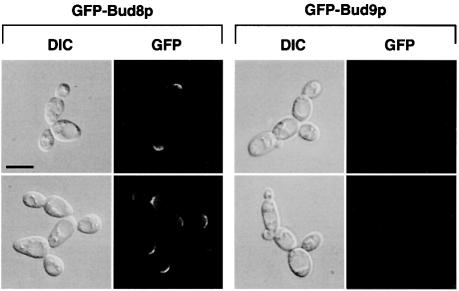
Fig. 7. Subcellular localization of GFP–Bud8p and GFP–Bud9p in living PH cells. Representative cells of wild-type strain RH2447 expressing either GFP–Bud8p from plasmid pME1772 or GFP–Bud9p from plasmid pME1777. Saccharomyces cerevisiae strains were grown in low ammonium media (SLAD/LA) for 15 h. Living cells were viewed by DIC or by fluorescence microscopy (GFP). Scale bar represents 5 µm.

An official website of the United States government
Here's how you know
Official websites use .gov
A
.gov website belongs to an official
government organization in the United States.
Secure .gov websites use HTTPS
A lock (
) or https:// means you've safely
connected to the .gov website. Share sensitive
information only on official, secure websites.
