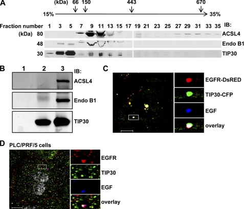
FIGURE 2.
Endogenous TIP30, ACSL4, and Endo B1 associate together. A, endogenous TIP30, ACSL4, and Endo B1 were co-sedimented in a glycerol gradient. Whole cell extracts of PLC/PRF/5 cells and protein markers were loaded on the top of linear 10–35% (v/v) glycerol gradients (10 ml) formed from the bottom of 12-ml Beckman tubes. The buffer throughout the gradients was 20 mm Hepes-KOH (pH 7.0), 100 mm KCl, 1 mm dithiothreitol. The gradients were centrifuged at 200,000 × g in a SW41 Ti rotor (Beckman) at 4 °C for 20 h. After centrifugation, fractions were collected, concentrated, and subjected to immunoblot (IB) analysis. Albumin (66 kDa), alcohol dehydrogenase (150 kDa) apoferritin (443 kDa), thyroglobulin (670 kDa), and blue dextran (2,000 kDa) were run in parallel as molecular mass indicators. B, direct interaction of TIP30, ACSL4, and Endo B1. GST-ACSL4 and His-Endo B1 (lane 1), FLAG-TIP30 (lane 2), or GST-ACSL4, His-Endo B1, and FLAG-TIP30 (lane 3) were subjected to anti-FLAG M2 immunoprecipitation. 100 ng of each protein was used. Aliquots of precipitates were resolved on SDS-PAGE and analyzed by immunoblot with the indicated antibodies. Purified FLAG-TIP30 was described previously (30). Bacterially expressed GST-ACSL4 was purchased from Abnova. His-Endo B1 was expressed in BL21. C, TIP30 and EGF-EGFR are colocalized in endosomes in response to EGF treatment. HepG2 cells co-expressing TIP30-CFP (green) and EGFR-DsRed (red) were treated with 10 ng/ml Alexa647-EGF (blue) for 10 min and then analyzed by confocal microscopy. A typical image is shown. The boxed areas are magnified. Scale bar, 10 μm. D, TIP30 is localized to EGFR-positive endosomes. PLC/PRF/5 cells were immunostained with antibodies for EGFR (red) and TIP30 (green) after 30 min of Alexa488-EGF (blue) internalization. The boxed areas are magnified. Scale bar, 10 μm.

