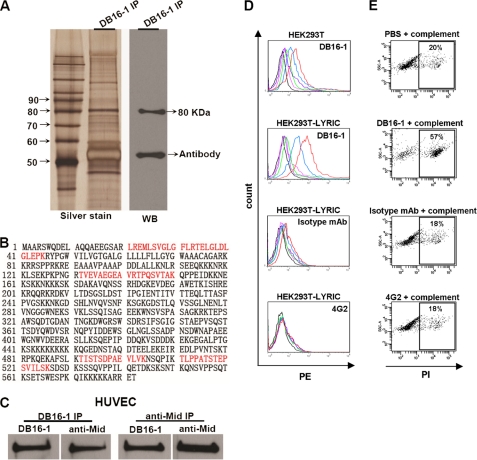
FIGURE 3.
Identification of the target protein of DB16-1 from HUVECs. A, the target protein was immunoprecipitated from HUVEC lysate by DB16-1 and analyzed by silver staining and Western blotting. B, peptide sequences of the target protein through LC-MS/MS analysis are marked. The peptide in red highlights the sequences present in LYRIC protein. C, co-immunoprecipitation (IP) of HUVEC lysates with DB16-1 antibody and a commercial polyclonal anti-LYRIC (anti-Mid) antibody and subsequent immunoblotting with anti-Mid and DB16-1 antibodies. These two antibodies recognized the same target in HUVECs. D, HEK293T cells expressing full-length LYRIC were analyzed by flow cytometry. DB16-1 bound to LYRIC-expressed HEK293T cells in a dose-dependent manner. Isotype mAb and 4G2 antibodies were used as negative controls. Red line, 50 μg/ml; blue line, 12.5 μg/ml; green line, 3.1 μg/ml; purple line, 0.78 μg/ml; pink line, 0.19 μg/ml; black line, 0 μg/ml. E, complement-dependent cytotoxicity assay was performed by flow cytometry in LYRIC-expressed HEK293T cells treated with complement alone, DB16-1 (50 μg/ml) plus complement, isotype mAb (50 μg/ml) plus complement, and 4G2 (50 μg/ml) plus complement.

