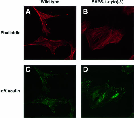
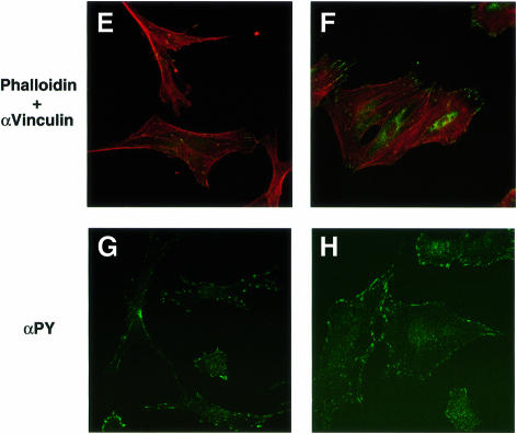
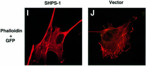
Fig. 2. Cytoskeletal architecture of wild-type and SHPS-1 mutant cells. Wild-type (A, C, E and G), SHPS-1–cyto(–/–) (B, D, F and H) or SHPS-1–cyto(–/–) cells transiently transfected with pTracer CMV containing (SHPS-1) (I) or not containing (vector) (J) SHPS-1 cDNA were seeded on glass coverslips and subsequently fixed and stained with rhodamine-labeled phalloidin (A, B, E, F, I and J). Cells were also stained with a mAb to vinculin (αvinculin) (C–F), and the distribution of phosphotyrosyl proteins was examined with mAb PY20 to phospho tyrosine (αPY) (G and H). Transfected cells were also detected by autofluorescence of green fluorescent protein (GFP) encoded by the vector (I and J). Cells were examined with a confocal microscope (original magnification, 630×).

An official website of the United States government
Here's how you know
Official websites use .gov
A
.gov website belongs to an official
government organization in the United States.
Secure .gov websites use HTTPS
A lock (
) or https:// means you've safely
connected to the .gov website. Share sensitive
information only on official, secure websites.


