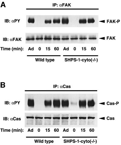
Fig. 5. Adhesion-induced tyrosine phosphorylation of FAK (A) and p130Cas (B) in wild-type and SHPS-1 mutant cells. Wild-type and SHPS-1–cyto(–/–) cells were either maintained adherent (Ad) or detached from culture dishes, replated on dishes coated with fibronectin (10 µg/ml) and incubated for the indicated times at 37°C. Cell lysates were prepared and subjected to immunoprecipitation (IP) with mAbs to FAK (αFAK) (A) or p130Cas (αCas) (B). Both types of immuno precipitate were then subjected to immunoblot analysis (IB) with horseradish peroxidase-conjugated mAb PY20 to phosphotyrosine (αPY). Duplicate immunoprecipitates were probed with polyclonal antibodies to FAK (αFAK) (A) or p130Cas (αCas) (B), as indicated, to verify the presence of equal amounts of FAK or p130Cas in each sample. The positions of FAK, tyrosine-phosphorylated FAK (FAK-P), p130Cas (Cas) and tyrosine-phosphorylated p130Cas (Cas-P) are indicated.

An official website of the United States government
Here's how you know
Official websites use .gov
A
.gov website belongs to an official
government organization in the United States.
Secure .gov websites use HTTPS
A lock (
) or https:// means you've safely
connected to the .gov website. Share sensitive
information only on official, secure websites.
