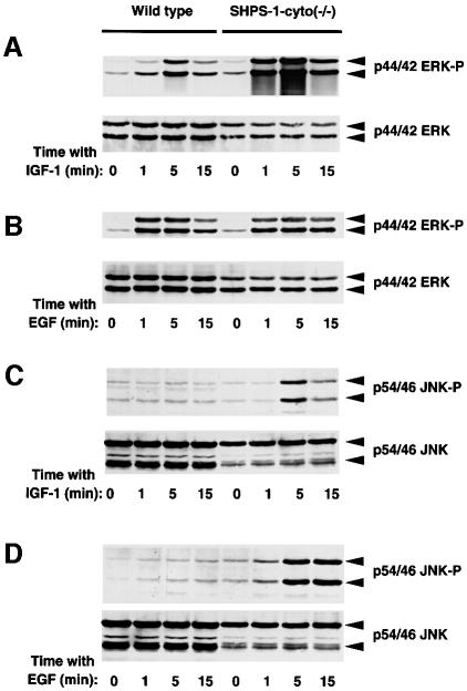
Fig. 8. Effects of SHPS-1 truncation on growth factor-induced activation of ERKs (A and B) and JNKs (C and D). Wild-type and SHPS-1–cyto(–/–) cells were deprived of serum for 18 h and then incubated in the presence of IGF-1 (100 ng/ml) (A and C) or EGF (100 ng/ml) (B and D) for the indicated times. Cell lysates were subjected to immunoblot analysis with antibodies specific for the activated (phosphorylated) forms either of p44 and p42 ERK (A and B, upper panels) or of p54 and p46 JNK (C and D, upper panels). Duplicate samples were also probed with antibodies to total ERK protein (A and B, lower panels) or to total JNK protein (C and D, lower panels). The positions of ERKs and JNKs, as well as of their phosphorylated forms, are indicated.

An official website of the United States government
Here's how you know
Official websites use .gov
A
.gov website belongs to an official
government organization in the United States.
Secure .gov websites use HTTPS
A lock (
) or https:// means you've safely
connected to the .gov website. Share sensitive
information only on official, secure websites.
