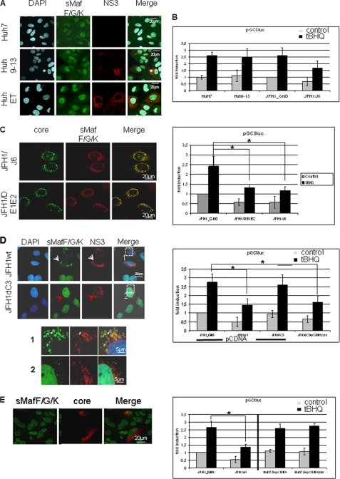
FIGURE 5.
HCV-dependent inhibition of Nrf2/ARE-regulated genes depends on core. A, confocal immunofluorescence microscopy (×630 magnification) of the cell lines Huh9–13 or HuhEtneo carrying the subgenomic replicons. HCV-negative cells (Huh7.5) served as control. Immunofluorescence staining was performed using the polyclonal sMaf-specific serum (green fluorescence) and an NS3-specific antibody (red). B, reporter gene assay of HuH9–13, Huh7.5, Huh7.5JFH1/J6, and Huh7.5/JFH1_GND cells transfected with the luciferase reporter construct harboring the GCS-derived ARE. The bars represent the standard deviation. *, p < 0.05. C, confocal immunofluorescence microscopy of Huh7.5 cells replicating the complete HCV genome (JFH1/J6) or the E1/E2 deletion construct (JFH1DE1/E2). Immunofluorescence staining was performed using the polyclonal sMaf-specific serum (red) and a core-specific antibody (green). The corresponding reporter gene assay was performed using the luciferase reporter construct harboring the GCS-derived ARE sequences. The bars represent the standard deviation. *, p < 0.05. D, confocal immunofluorescence microscopy of Huh7.5 cells replicating the complete HCV genome (JFH1wt) or the core deletion construct (JFH1dC3) Immunofluorescence staining was performed using the polyclonal sMaf-specific serum (green) and a NS3-specific antibody (red). Higher magnification of the indicated fields is shown in 1 and 2. The corresponding reporter gene assay of JFH1wt- or JFH1dC3-replicating HuH7.5 cells is shown below. 48 h after electroporation cells were co-transfected with the reporter constructs harboring the GCS-derived ARE sequences and pCDNA3 vector as control. Co-transfection with pCDNAcore that encodes HCV core rescues the inhibitory effect of the core-deficient replicon construct. The bars represent the standard deviation. *, p < 0.05. E, confocal immunofluorescence microscopy of Huh7.5 cells that selectively overexpress HCV core after transient transfection with a core expression vector. Immunofluorescence staining was performed using the polyclonal small Maf-specific serum (green) and a core-specific antibody (red). Corresponding reporter gene assay of Huh7.5 cells that were co-transfected with pCDNAcore and the reporter constructs harboring the GCS-derived ARE sequences is shown below. Transfection with pCDNA3 vector or cotransfection of JFH1wt or JFH1-GND cells served as controls. The bars represent the standard deviation. *, p < 0.05. A–E, cells were stimulated with tert-butylhydroquinone (tBHQ) (black bars) or left untreated (gray bars).

