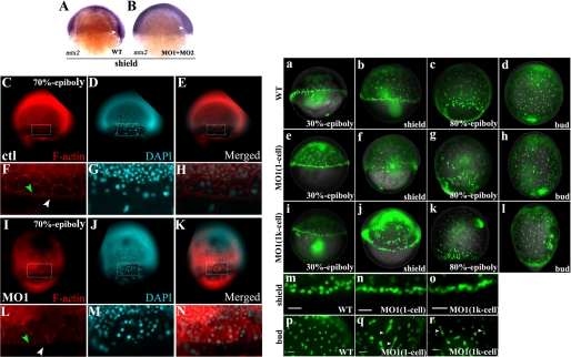
FIGURE 4.
mtx2 expression and the F-actin ring are altered in apoa2 morphants and YSN are disordered in the absence of apoa2 function. A, mtx2 expressed as YSL-specific gene expressed blastoderm margin at shield stage. B, mtx2 expression was decreased significantly with loss of apoa2. White arrowheads indicate the blastoderm. C–N, F-actin antibody and DAPI-stained embryos pre-injection with ctl MO or apoa2 MO1 at the 1-cell stage. F–H and L–N denotes magnified views of the regions marked by rectangles in C–E and I–K. The F-actin punctate band of the eYSL (indicated with white arrowhead) and the peripheral F-actin ring within the EVL (indicated with green arrowhead) are shown. Wild-type (a–d and m, p) and apoa2 MO1 (injection at 1-cell) (e–h and n, q), apoa2 MO1 (injection at 1k-cell) (i–l and o, r) embryos were injected with SYTOX Green at the 1k-cell stage, then observed and imaged at 30%-epiboly, shield, 80%-epiboly, and tail-bud stages. The YSN at blastoderm margin exhibited abnormal aggregation in the apoa2 losing embryos (n and q) compared with wild-type (m) at the shield stage. YSN were regularly spaced in wild-type (p); but random distribution in apoa2 MO1 (q and r). The white arrowhead indicates incomplete dissociation of YSN. Scale bar, 50 μm. (b–c, f–g, and j–k) show lateral views and (d, h, and l) show dorsal views.

