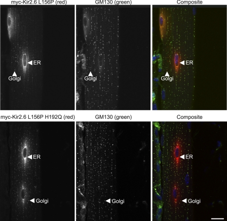
FIGURE 8.
Kir2.6-L156P is trafficked to Golgi following electroporation in mouse skeletal muscle. Mouse tibialis cranialis skeletal muscle fibers were electroporated in vivo with Myc-Kir2.6-L156P or Myc-Kir2.6-L156P/H192Q. Seven days later, tissue was fixed and labeled with anti-Myc (red), anti-GM130 (green), and DAPI (blue). The Kir2.6-L156P mutant is trafficked out of the ER to the Golgi (arrowhead), and trafficking is enhanced in the double mutant Kir2.6-L156P/H192Q. Bar, 20 μm.

