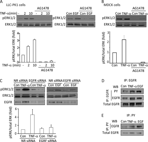
FIGURE 1.
Inhibition of the EGFR prevents TNF-α-induced ERK activation. A and B, LLC-PK1 (A) or MDCK (B) cells were grown to confluence and serum-depleted for 3 h before the treatments. The cells were pretreated with 100 nm AG1478 for 30 min in serum-free DMEM followed by the addition of 10 ng/ml TNF-α for 2 or 10 min (A) or 10 min (B) or 100 ng/ml EGF for 10 min as indicated. Where used, the inhibitor was present throughout the TNF-α or EGF treatment. At the end of the treatments the cells were lysed, and pERK and ERK were detected using Western blotting. C, MDCK cells were transfected with NR siRNA or an siRNA against canine EGFR. Forty-eight hours after transfection the cells were serum-depleted and then treated with 10 ng/ml TNF-α (5 min) or 100 ng/ml EGF (10 min). ERK and pERK were detected as above. The graphs below the blots summarize densitometric analysis of the pERK blots (see “Experimental Procedures”). Data presented are the mean ± S.E. of n = 3 experiments. D and E, LLC-PK1 cells were transfected with human EGFR. 48 h later the cells were serum-depleted and treated with 10 ng/ml TNF-α (15 min) or 10 ng/ml EGF (5 min). At the end of the treatment cells were lysed and immunoprecipitated (IP) using EGFR (D) or phosphotyrosine (PY) (E) antibody as described under “Experimental Procedures.” The immunoprecipitated proteins and samples of the total cell lysates were analyzed by Western blotting (WB) using Tyr(P) and EGFR antibodies as indicated. In D the transfected EGFR in the total cell lysates was also visualized. The EGFR antibody in the total cell lysates and precipitates from untransfected cells yielded only a marginal signal (not shown).

