FIGURE 5.
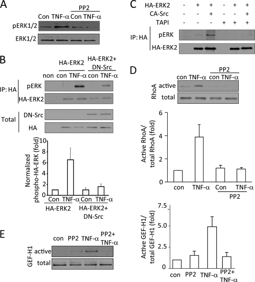
Src kinase mediates TNF-α-induced ERK, GEF-H1, and RhoA activation. A, D, and E, LLC-PK1 cells were pretreated with 10 μm Src family inhibitor PP2 (15 min) followed by 10 ng/ml TNF-α for 5 min. The inhibitor was present during the TNF-α treatment. Levels of pERK (A), RhoA (D), or GEF-H1 (E) were determined as described earlier. B and C, LLC-PK1 cells were transfected with HA-tagged ERK2 with or without dominant negative Src kinase (B) or active Src kinase (C). Forty-eight hours after transfection the cells were treated with TNF-α (10 ng/ml, 10 min) as indicated. In C, cells were pretreated for 1 h with TAPI-1 as indicated before the addition of TNF-α. At the end of the treatment the cells were lysed, and HA-ERK2 was immunoprecipitated (IP). Phospho-ERK was detected in the precipitates. The blots were stripped and redeveloped using an HA-antibody to detect precipitated HA-ERK2. In B, the transfected HA-ERK2 and avian dominant negative Src were also detected in the total cell lysates. Please note that the avian Src antibody does not react with mammalian Src. In the experiments shown on C neither the transfected HA-ERK nor the avian Src was detectable in the total cell lysates (not shown). No signal was visible when untransfected cell lysates are used (non). The graph in B shows densitometric analysis done as described under “Experimental Procedure” (mean ± S.E. n = 3).

