
FIGURE 5.
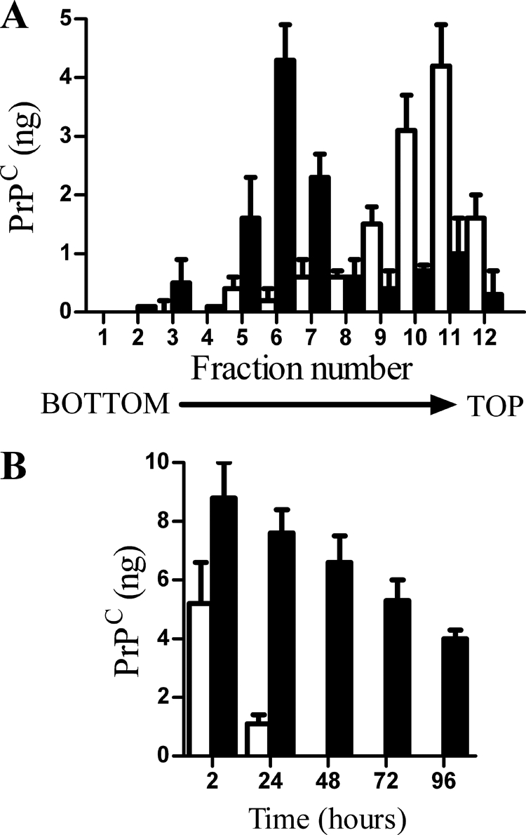
Acylation of GPI anchors affected the targeting of PrPC to lipid rafts. A, PrP null cortical neurons were incubated with 10 ng of PrPC-GPI (□) or PrPC-G-lyso-PI (■) for 2 h. Cell extracts were prepared and separated by centrifugation on a sucrose density gradient, and the amount of PrPC in each fraction was determined by ELISA. Values shown are the mean amount of PrPC (ng) ± S.D. from triplicate experiments performed three times (n = 9). B, PrP null cortical neurons were pulsed with 10 ng of PrPC-GPI (□) or PrPC-G-lyso-PI (■) for 2 h. The amount of PrPC in cells was determined at time points thereafter as shown. Values shown are the mean amount of PrPC (ng/106 cells) ± S.D. from triplicate experiments performed four times (n = 12).