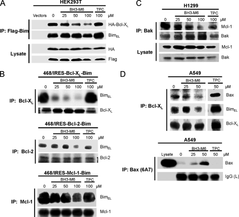
FIGURE 4.
BH3-M6 inhibits the interaction between Bcl-XL, Bcl-2, and Mcl-1 with Bim, Bak, or Bax in intact cells. A, co-immunoprecipitation from HEK293T cells. HEK293T cells were co-transfected with HA-Bcl-XL and Flag-BimEL for 18 h. Cells were exposed to 0, 25, 50, and 100 μm of BH3-M6 or 100 μm of TPC for 2 h at 37 °C, lysed, and subjected to immunoprecipitation with anti-FLAG-M2 beads. The resulting immune complexes, as well as total lysates, were analyzed by Western blotting with the indicated antibodies. B, co-immunoprecipitation from MDA-MB-468 cells expressing Bcl-XL-IRES-BimEL, Bcl-2-IRES-BimEL, and Mcl-1-IRES-BimEL. Cells were grown in 100-mm plates and treated with 0, 25, 50, and 100 μm BH3-M6 or 100 μm TPC for 24 h at 37 °C, lysed, and subjected to immunoprecipitation with Bcl-XL, Bcl-2, and Mcl-1 antibodies. The resulting immune complexes were analyzed by Western blotting with Bim, Bcl-XL, Bcl-2, and Mcl-1 antibodies. C, co-immunoprecipitation from H1299 cells. H1299 non-small lung cancer cells were grown in RPMI 1640 medium plus 10% FBS, antibiotics and 1% sodium pyruvate, 1% HEPES, and 1.1% glucose. They were seeded in 100-mm plates and treated with 0, 25, 50, and 100 μm BH3-M6 or 100 μm TPC for 24 h at 37 °C, lysed and subjected to immunoprecipitation with Bak antibody. The resulting immune complexes were analyzed by Western blotting with the indicated antibodies. D, co-immunoprecipitation from A549 cells. A549 cells were grown in F-12K medium plus 10% FBS and antibiotics and then serum-starved for 20 h, followed by treatment with 0, 25, and 50 μm of BH3-M6 or 50 μm of TPC for 1 h at 37 °C. Cells were then lysed and subjected to immunoprecipitation with Bcl-XL antibody (upper panel) and Bax 6A7 antibody (lower panel). The resulting immune complexes were analyzed by Western blotting with the antibodies indicated on the right.

