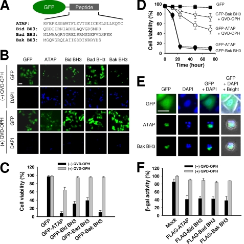
FIGURE 1.
Transient expression of GFP-ATAP or GFP-BH3 peptides induces apoptosis of HeLa cells. A, schematic diagram of constructs expressing ATAP or BH3 peptides fused with GFP protein. B and C, HeLa cells were transfected with 1 μg of plasmids expressing GFP-ATAP- or GFP-BH3-peptide fusion proteins in the absence or presence of the pan-caspase inhibitor QVD-OPH (50 μm). At 24 h after transfection, cells were stained with DAPI and observed under a fluorescence microscope. B, representative photographs of GFP- or DAPI-positive cells transfected with indicated plasmids. The two images in each column were from the same field. Bar, 10 μm. C, percentage of surviving cells was determined by the ratio of GFP-positive cells without DAPI staining to total GFP-positive cells. About 200 cells from three different fields were scored. Data are expressed as the means ± S.E. D, time course effect of GFP-ATAP and GFP-Bak BH3 on HeLa cells. Cell survival was measured by DAPI exclusion in the HeLa cells transfected with 1 μg of plasmids expressing GFP-ATAP or GFP-Bak BH3. E, transient expression of GFP-ATAP- or GFP-Bak BH3-induced apoptotic nuclear morphology. HeLa cells were transfected with GFP-ATAP or GFP-Bak BH3. At 24 h of transfection, cells were fixed, stained with DAPI, and observed under a fluorescence microscope. Bar, 10 μm. F, cytotoxic activity of FLAG-tagged ATAP or BH3 peptides. HeLa cells were co-transfected with 1 μg of the indicated plasmid expressing FLAG peptide and 0.1 μg of pCMV-β reporter plasmid either in the presence or absence of 50 μm QVD-OPH. At 24 h after transfection, β-galactosidase activity was measured. Cell viability is shown as the relative β-galactosidase activity of the FLAG peptide-expressing cells to the control cells.

