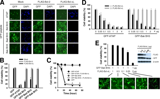
FIGURE 2.
Bcl-xL or Bcl-2 does not inhibit ATAP-induced apoptosis but efficiently inhibits BH3 peptide-induced apoptosis. A and B, HeLa cells were co-transfected with 1 μg of plasmids expressing GFP-ATAP or GFP-BH3 peptides along with 1 μg of mock vector, pFLAG-Bcl-2, or pFLAG-Bcl-xL. At 24 h after transfection, cells were treated with DAPI and observed under a fluorescence microscope. A, representative photographs show GFP- or DAPI-positive cells in the same field. Bar, 10 μm. B, percentage of surviving cells was determined by the ratio of GFP-positive cells without DAPI staining to total GFP-positive cells. About 200 cells from three different fields were scored. Data are expressed as the means ± S.E. C, time course effect of FLAG-Bcl-xL on the apoptosis induced by GFP-ATAP or GFP-Bak BH3 in HeLa cells. Cell survival was measured by DAPI exclusion in the HeLa cells transfected with 1 μg of plasmids expressing GFP-ATAP or GFP-Bak BH3 with or without 1 μg of FLAG-Bcl-xL plasmid. D, HeLa cells were transiently co-transfected with 1 μg of FLAG-Bcl-xL plasmid and various amounts of GFP-ATAP or GFP-Bak BH3 construct as indicated. The percentage of surviving cells was determined by DAPI exclusion assay as described previously above. Data are expressed as the means ± S.E. E, HeLa cells were transiently co-transfected with 1 μg of GFP-Bak BH3 construct and various amounts of FLAG-Bcl-xL construct as indicated. The percentage of surviving cells was determined by the DAPI exclusion assay as described above. Data are expressed as the means ± S.E. Right panel, protein expression in the transfected cells was assessed by immunoblotting the cell lysates using antibodies specific for FLAG, GFP, or β-actin. Lower panel, representative photographs show cellular distribution of GFP-Bak BH3 fusion protein in the transfected cells. Bar, 10 μm.

