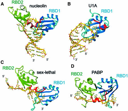
Fig. 7. Comparison between nucleolin RBD12–sNRE complex and the other RBD–RNA complexes. (A) Nucleolin RBD12–sNRE complex. (B) U1A RBD1 bound to U1 snRNA stem–loop II (Oubridge et al., 1994). (C) Sex-lethal RBD12–UGU8 complex (Handa et al., 1999). (D) PABP RBD12–A8 complex (Deo et al., 1999). Note that the location of the amino acids on the surface of the β-sheet varies among the different RBDs. In all panels, RBD1 is shown as a ribbon in cyan and RBD2 is in green. The RNA is in yellow, represented as sticks.

An official website of the United States government
Here's how you know
Official websites use .gov
A
.gov website belongs to an official
government organization in the United States.
Secure .gov websites use HTTPS
A lock (
) or https:// means you've safely
connected to the .gov website. Share sensitive
information only on official, secure websites.
