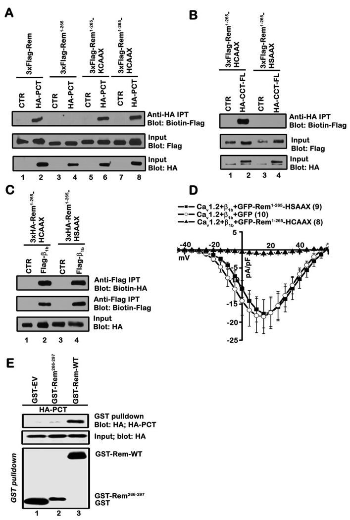
Figure 4. Plasma membrane targeting is necessary for Rem:CCT association.
A, TsA201 cells were transiently co-transfected with the indicated Rem and CCT expression vectors. Co-immunoprecipitation was performed with anti-HA antibody and interaction with Rem examined by immunoblotting with biotinylated FLAG antibody. Results are representative of four independent experiments. B, TsA201 cells were transiently co-transfected with the indicated plasmids and co-immunoprecipitation performed with HA antibody as described in A. Results are representative of three independent experiments. C, TsA201 cells were transfected with the indicated plasmids. Co-immunoprecipitation was performed with Flag antibody and interaction with Rem proteins examined by immunoblotting with biotinylated anti-HA antibody. Immunoprecipitates were blotted for β1b using biotinylated Flag antibody. Results are representative of three independent experiments. D, TsA201 cells were transfected with plasmids expressing Cav1.2, β1b and either GFP-Rem-(1-265)-HCAAX, GFP-Rem-(1-265)-HSAAX, or unfused GFP as control. Current was examined using the whole-cell patch clamp configuration in the presence of 30 mM Ba2+. E, TsA201 cells were transiently co-transfected with GST alone, GST-Rem, or GST-Rem(266–297) and PCT. GST fusion proteins were isolated using glutathione-Sepharose resin and interaction with PCT examined by immunoblotting. Results are representative of three independent experiments.

