
Figure 2.
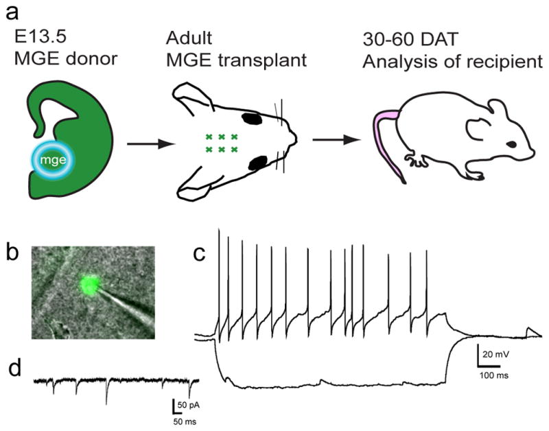
Following adult hippocampal transplantation, GFP+ graft-derived cells become functional neurons. A. An adult CD-1 mouse (P101) received a GFP+ MGE cell graft and was sacrificed 47 DAT for electrophysiological analysis. B. Image of GFP+ cell for which the recordings are shown in C and D. C. Cell fired action potentials with fast afterhyperpolarizations that are characteristic of interneurons. Excitatory postsynaptic potentials (EPSPs) can be seen during and after the hyperpolarizing current step. D. Use of an internal solution containing KGluconate and KCl (Bacci et al., 2003) allowed voltage clamp recordings from the same cell. Spontaneous inhibitory postsynaptic currents (IPSCs) recorded in the presence of glutamate receptor blockers demonstrate that GFP+ cells receive inhibitory synaptic input. Vhold = −60 mV.