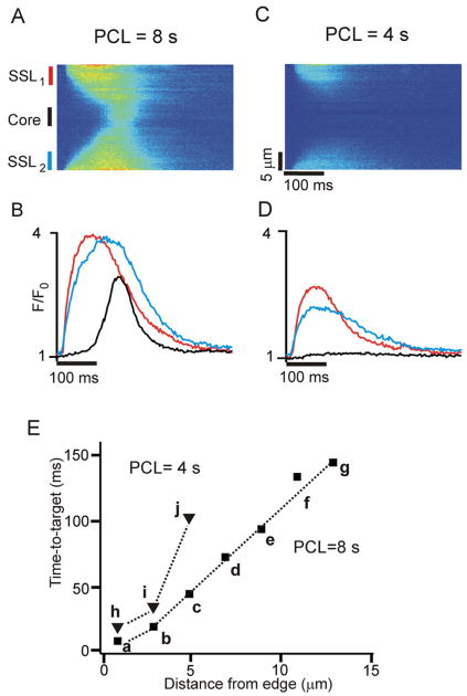
Figure 1.
Ca2+ waves occur at very low pacing rates; local subsarcolemmal (SSL) elevations in Ca2+ occur at higher pacing rates. (A) Space-time line scan image obtained at PCL = 8 s shows an early increase in [Ca2+] in SSL regions and a delayed increase in the core. (B) Local Ca2+ transients averaged over 5 μm at two SSL regions (red and blue) and in cell core (black), as indicated to the left of the image in (A). (C) and (D) Space-time image and local Ca2+ transients, respectively, obtained in the same cell at PCL = 4 s. Electrical stimulation induces elevations in Ca2+ only in the SSL regions. (E) Time-to-target plot of activation time versus distance from cell edge, with Ca2+ transients averaged over each 2 μm region.

