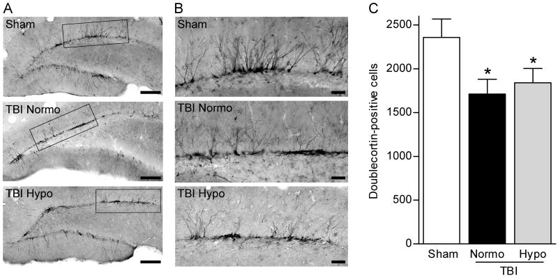
Fig. 4.
Numbers of doublecortin-positive cells are decreased at chronic time points after brain injury and this decrease is not rescued by hypothermia therapy. Low magnification images (20X) of doublecortin immunostaining of the dentate gyrus at 12 weeks post-TBI (A). Higher magnification (60X) of doublecortin-positive cells revealed that the dendritic branches were more laterally oriented in the injured hippocampus as compared to the non-injured hippocampus (B). Quantification by stereology revealed that numbers of doublecortin-positive cells were significantly decreased in both TBI-normothermic (n=12) and TBI-hypothermic animals (n=16) as compared to sham animals (n=15) (C). *P<0.05 for sham versus TBI-normothermic animals or TBI-hypothermic animals. Data represent mean ± SEM. (A) Scale bars, 200 μm. (B) Scale bars, 50 μm.

