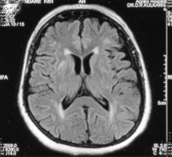
Figure 1.

T2 weighted FLAIR image showing multiple white matter abnormalities (hyper intensities) at left frontal, bilateral parietal, occipital white matter. Also noted are sub-cortical grey matter hyper intensities in head of right caudate nucleus and bilateral lentiform nuclei.
