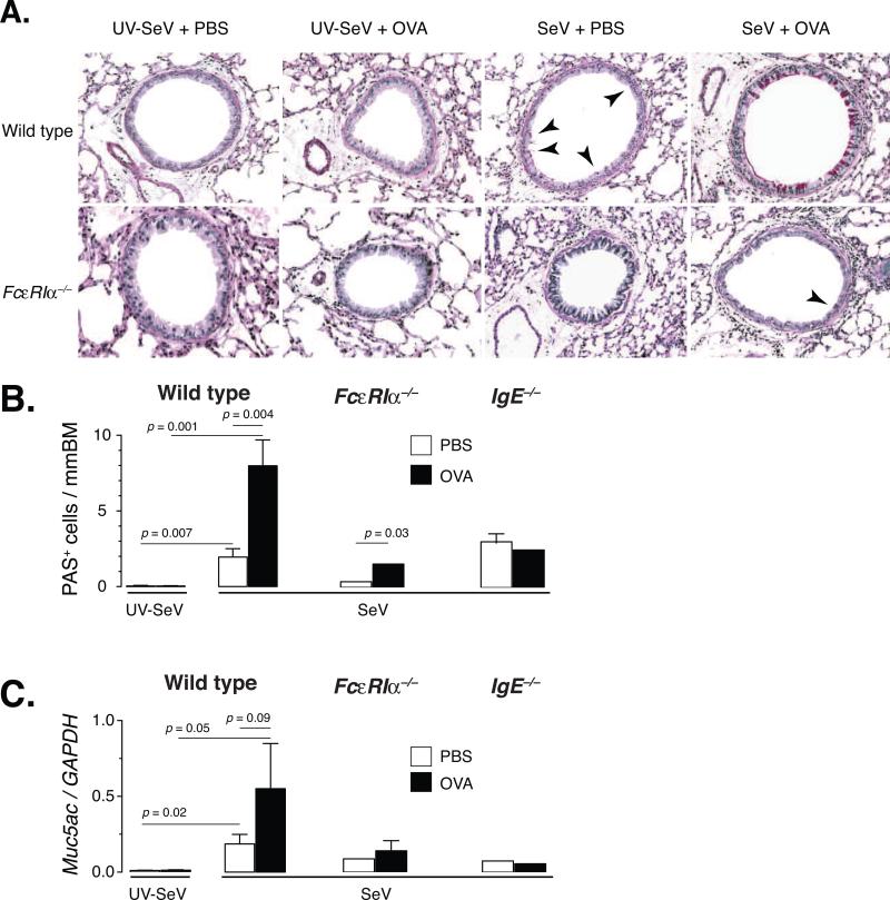
Figure 4. Non-viral antigen exposure during a severe paramyxoviral infection induces mucous cell metaplasia primarily through an FcεRI-dependent process.
(A) Representative PAS staining of lung sections from C57BL6 or FcεRIα–/– mice inoculated with either 2×10⁵ pfu SeV or UV-SeV i.n., exposed to OVA or PBS i.n. on day 8 p.i., and challenged with OVA i.n. daily as outlined in figure 1C. SeV infection induced a modest level of PAS positive cells (red staining cells in bronchial epithelium, indicated by arrowheads) in wild-type mice, but exposure to OVA during the viral infection greatly enhanced this response (the arrowheads were left off due to the much greater level of staining). Mice deficient in FcεRIα had very few PAS positive cells in their airways (see arrowhead), regardless of treatment. (B) Quantification of the number of PAS positive cells per mm BM in the indicated mouse strain inoculated with UV-SeV (wild-type only) or SeV and then given PBS (open bars) or OVA (filled bars) and treated as in (A). (C) Level of expression of Muc5ac gene product in whole lung (normalized to GAPDH levels; right graphs) for each of the groups as discussed in (B). Data are presented as mean +/- SEM, with the number of mice as outlined in the legend to figure 3. Statistical comparisons are as indicated using two-tailed, unpaired Student's t-test.

