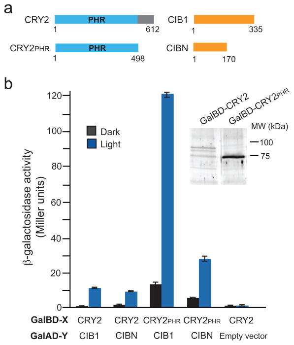
Figure 1.
Mapping of interacting domains of CRY2 and CIB1. (a) Schematic showing full-length CRY2 and CIB1 constructs used in experiments. The numbers below the proteins indicate amino acid residue position. (b)β-galactosidase activity of CRY2 and CIB1 constructs tested for interaction in the dark or in blue light (461 nm, 1.9 mW, 4 hr). The Gal4 binding domain (Gal4BD-X) and Gal4 activation domain (Gal4AD-Y) fusions used are indicated. The control vector was pGBKT7rec containing no insert. Error bars represent standard deviation (n = 3 samples). The inset panel shows immunoblot analysis of Gal4BD fusion proteins in yeast.

