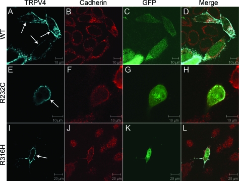
Figure 3. Localization of wild-type and mutant TRPV4 on the plasma membrane in HeLa cells.
Confocal microscopy was performed using HeLa cells transfected with plasmids pIRES2-ZsGreen1 containing wtTRPV4 (A–D), TRPV4R232C (E–H), and TRPV4R316H (I–L). Cells expressing exogenous TRPV4 were labeled by green fluorescent protein (GFP) (C, G, and K). TRPV4 is shown by blue (A, E, and I) and cadherin by red (B, F, and J). Merged images are shown on the right panels (D, H, and L). Arrows indicate TRPV4 signals on the plasma membrane.

