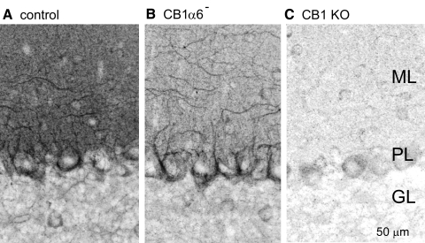
Fig. 1.
Immunostaining reveals selective elimination of CB1Rs from cerebellar granule cells in CB1α6− animals. Sagittal sections (50 μm thick, ≥4 per animal) from 2-mo-old mice were stained with a rabbit polyclonal antibody raised against the last 15 amino acids of the CB1R C-terminus (Bodor et al. 2005; Nyiri et al. 2005) and imaged at ×40. Images (z-projections of 11 images taken at 1-μm intervals) of the cerebellar cortex are shown for a representative experiment of control (A), CB1α6− (B), and global CB1 knockout (C) animals (n = 5 each). The molecular layer (ML), the Purkinje cell layer (PL), and the granular layer (GL) are indicated in C.

