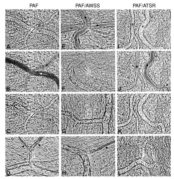
Figure 4.
Videomicrographs showing inhibition of PAF-induced sickle erythrocytes adhesion in the ex vivo mesocecum microvasculature in the presence of ICAM-4 peptide ATSR. (A-D) Ex vivo preparation treated with PAF. (A) Clear vessel lumen (a = arteriole, v = venule) during perfusion with Ringer-albumin solution; (B and C) sickle erythrocytes bolus (arrows in B indicate flow direction) is followed by adhesion of these cells exclusively in the venule but not in the arteriole; (D) Maximal adhesion is observed in small-diameter postcapillaty venules (arrow-heads), frequently resulting in occlusion. (E-H) Ex vivo preparation treated with PAF and an ICAM-4 F strand control peptide AWSS; (E and F) Infusion of sickle erythrocytes bolus results in adhesion in the venules (v), but not in the arteriole (b); (G) adherent sickle erythrocytes in venules; (H) adhesion results in frequent blockage of small-diameter venules (arrow-head). (I-L) Ex vivo preparation treated with PAF and ICAM-4 peptide ATSR; (I) clear vessel lumens during perfusion with Ringer-albumin; (J and K) after a bolus infusion of sickle erythrocytes, rapid flow is observed in both arteriole and venules (arrows indicate flow direction), resulting in no adhesion; (L) scanning of the vasculature revealed little or no adhesion in venules. From Kaul et al. (50).

