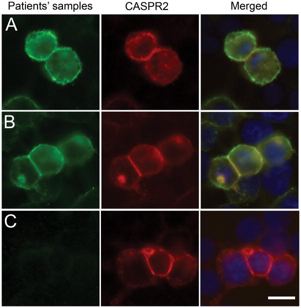
Figure 1. Patients’ antibodies react with cells expressing Caspr2.
HEK cells were transiently transfected to express human Caspr2, and labeled with the index patient’s CSF (A; green) or serum from a different patient (B; green) and a rabbit antibody to Caspr2 (A, B red), and counterstained with DAPI. Merged images (A, B yellow) demonstrate the overlap of patients’ antibody staining with Caspr2 expression. CSF (C) from a control patient did not react with cells expressing Caspr2. Scale bar: 20 μm

