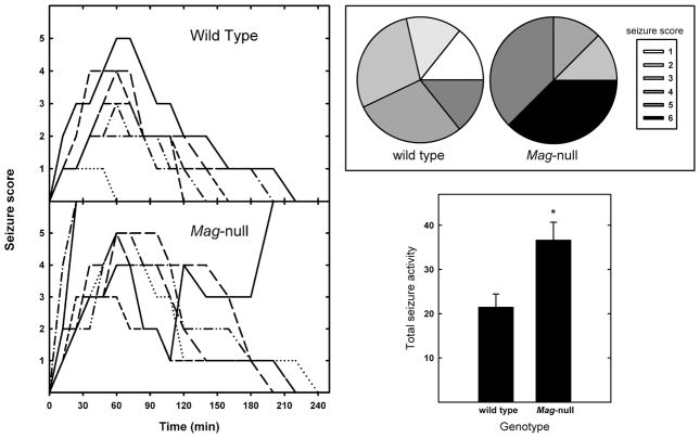
Fig. 1.
Mag-null mice display increased susceptibility to kainic acid-induced seizures. (Left) Seizures were induced in WT (n = 7) and Mag-null mice (n = 8) by intraperitoneal injection of KA (25 mg/kg). Seizure activity was video recorded and quantified as follows: 0, normal behavior; 1, ceases exploring, grooming and sniffing (becomes motionless); 2 forelimb and/or tail extension, rigid posture; 3, myoclonic jerks of the head and neck with brief twitching or repetitive movements, head bobbing or “wet-dog shakes”; 4, forelimb clonus and partial rearing and falling; 5, forelimb clonus, continuous rearing and falling; 6 tonic-clonic movements, loss of posture. Mice that died due to seizure were scored at the highest level for the remainder of the observation period. Each trace represents an individual mouse. The traces represent seizure activity recorded every 12–20 min over a 4-h observation period. (Upper Right) Pie charts indicate the percent of mice reaching the indicated maximum seizure score for each geneotype. (Lower Right) Total seizure activity (mean ± SEM) is presented as the sum of the seizure scores at each time analyzed. Mag-null mice displayed significantly greater seizure activity (*, p <0.02).

