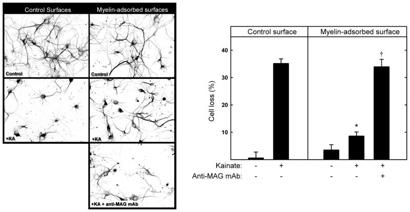
Fig. 4.
MAG protects hippocampal neurons from kainic acid-induced excitotoxicity in vitro. (Left) Hippocampal neurons were plated onto control surfaces or the same surfaces adsorbed with detergent-extracted proteins from rat brain myelin. As indicated, 1 h after plating, some cultures were treated with 10 μg/ml anti-MAG mAb. After 48 h, the medium was replaced with fresh control medium or medium containing 130 μM kainic acid (KA) to induce excitotoxicity. After an additional 24 h, the cultures were incubated for 30 min with medium containing propidium iodide, and then were fixed and stained with anti-tubulin mAb (TUJ1). Representative fluorescent micrographs are presented as reverse gray scale images to enhance clarity. (Right) Cell survival (mean ± SEM) was quantified (as described in the text) and normalized with respect to control surfaces. Live cell counts from 4 microscopic fields from each of 9 microwells from three independent experiments were averaged. Growth on myelin-adsorbed surfaces provided significant neuroprotection from KA (*, p < 0.001) compared to cells grown on control surfaces. Addition of anti-MAG antibody reversed this protection, resulting in cell loss that was not significantly different from KA-treated cells grown on control surfaces (†, p > 0.7).

