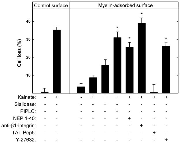
Fig. 7.
MAG-mediated protection from kainic acid-induced excitotoxicity is predominantly via NgR and β1-integrin. Hippocampal neurons were plated on control surfaces or the same surfaces adsorbed with detergent-extracted rat myelin. As indicated, 1 h after plating the following agents were added to the cultures: 10 mU/ml sialidase, 0.5 U/ml PI-PLC, 1 μM NEP1–40, 2.5 μg/ml function-blocking anti-β1-integrin receptor mAb, 100 nM TAT-Pep5, or 5 μM Y-27632. After 48 h, the media were replaced with fresh media containing the same agents plus 130 μM KA. After an additional 24 h, the cultures were incubated for 30 min with medium containing propidium iodide and then fixed and stained with TUJ1 mAb. Cell survival (mean ± SEM) was quantified and normalized with respect to the control. Neurons cultured on myelin-adsorbed surfaces were significantly protected from KA-induced toxicity (see Fig. 4). Significant reversal of myelin-mediated protection was observed upon addition of PI-PLC, NEP1–40, anti-β1-integrin mAb, and the ROCK inhibitor Y-27632 (*, p < 0.001 compared to KA-treated cultures on myelin-adsorbed surfaces without added agents). Statistically significant reversal of protection was not observed upon treatment with sialidase or TAT-Pep5.

