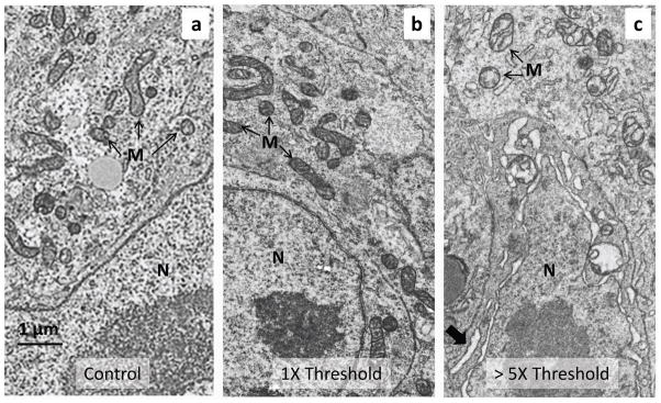
Figure 4. Transmission electron microscopy (TEM) after the OP procedure.
a-c, The TEM images show a typical cardiomyocyte in the inflow (sinoatrial) region of the heart tube of three different quail embryos, where laser light was directed. Each embryo was between 52 to 58 hours of development. The torso of each embryo was excised and fixed immediately after OP. a, Control embryo. The embryo was exposed to the OP experimental procedure, except that the pacing laser was not turned on during the experiment. b, Embryo paced slightly above threshold. The embryo heart was paced for approximately 20 seconds with 2 ms pulses (2.64 mJ/pulse) at 2 Hz. c, Embryo paced well above threshold. The embryo heart was paced for approximately 20 seconds with 4 ms pulses (13 mJ/pulse) at 2 Hz. The micrograph image was taken from a border region separating severely damaged tissue (partially ablated) from apparently healthy tissue. The mitochondria are vacuolated and the nuclear envelope is swollen, as are elements of the rough ER (large arrow). The cells and mitochondria in 4a and b have a similar appearance and show no signs of damage from the pulsed laser light. Some subtle abnormalities in the cristae present in cells from both threshold paced and unpaced control hearts are due to the unavoidable delay in excising the embryonic torso. N -nucleus, M - mitochondria.

