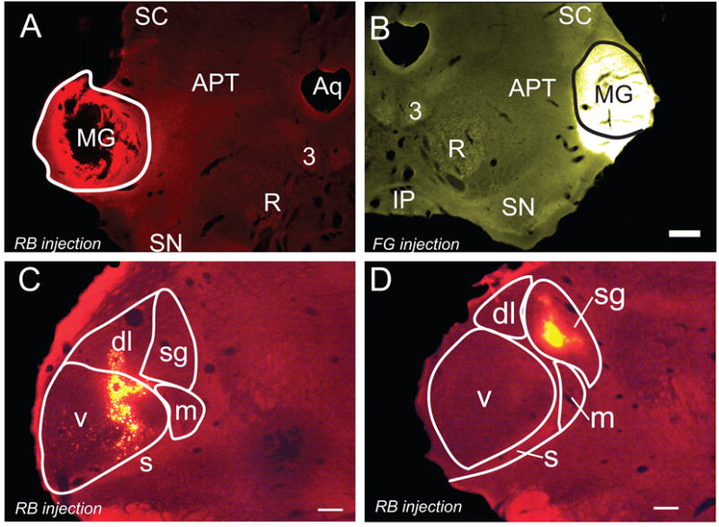
Figure 1.

Photomicrographs of representative large and small tracer injections into the MG. A) Large red bead (RB) injection that was confined to the left MG (solid outline). GP 633. The black area in the center of the injection represents an area in which the tissue fell loose during processing. B) A large FluoroGold injection that spread ventrally beyond the borders of the right MG. GP 633. Solid line - approximate borders of the MG. Scale bar applies to A and B (500 μm). C) Smaller injection of red beads that is centered in the ventral MG (v) and extends into the dorsolateral subdivision (dl). GP 595R. D) A small injection of red beads within the suprageniculate subdivision (sg). GP 586R. Transverse sections; dorsal is up; lateral is to the left in A, C-D and to the right in B. Scale bars = 500 μm. 3, oculomotor nucleus; APT, anterior pretectal nucleus; Aq, aqueduct; dl, dorsolateral; m, medial; s, shell; SC, superior colliculus; sg, suprageniculate; SN, substantia nigra; v, ventral. C and D adapted from Motts and Schofield, 2010, with permission.
