
Figure 3. JPH2 knock-down induces hypertrophy of HL-1 cells.
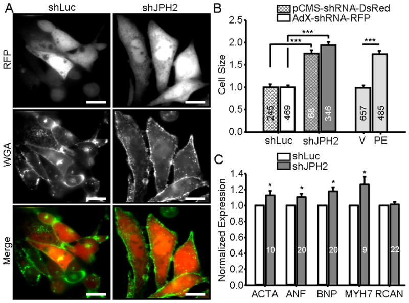
A. Representative image of RFP-positive HL-1 cells infected with control pCMS-shLuc or pCMS-shJPH2 and labeled with WGA. Scale bar, 100μm. B. Bar graph showing mean HL-1 cell size transiently transfected with pCMS-shRNA (cross-hatch) or adenovirally infected with AdX-shRNA (no cross-hatch). Delivery of shJPH2 by either transient transfection or adenoviral infection (dark gray bars) induced an increase in cell size compared to controls (white bars) that was not significantly different between oligo-delivery methodologies. Light gray bars, treatment with phenylephrine (PE), induced a similar increase in cell size compared to vehicle-treated controls (V). Numbers in bar graph indicate number of cells analyzed. ***, P < 0.0001. C. Relative RNA expression levels of ACTA, ANF, BNP, MYH7, RCAN1-4 in HL-1 cells infected with AdX-shJPH2-RFP normalized control cells. Numbers in bar graphs indicate number of assays. *, P < 0.05.