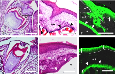
Fig. 4.
Light micrographs of the tooth germ and covering oral epithelium of rats of 10th day (a–c) and 15th (d–f) day after birth stained with H-E (a, b, d, e) or anti-Hsp27 antibody (c, f). Higher magnification of solid frames in a and d were showed in b and e, respectively. c and f represent similar areas to b and e, respectively. Basal membrane is indicated by dotted line (c, f). Hsp27-immunoreactivity was found in the prickle/granular and ameloblastic layers (arrowheads). The stellate reticulum (double asterisks in b, c) and papillary layer (double asterisks in e, f) as well as basal layer (arrows) were immunonegative. Single asterisk indicates the decalcified enamel. Bars=500 µm (a, d); 50 µm (b, c); 20 µm (e, f).

